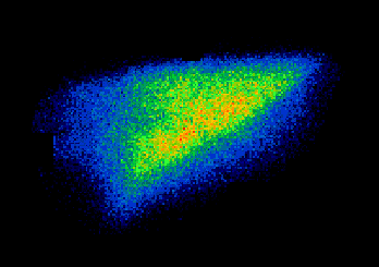
Figure 1: 99mTc aerosol ventilation image in horses. Ventilation image of the left lung acquired following inhalation of 99mTc-aerosol (left lung viewed from the side).
Because of progress in human medicine, the prevalence of numerous diseases has decreased. Unfortunately, asthma and COPD escape to the rule (Siafakas et al., 1989; Dubois et al., 1998). In the Laboratory for Functional Investigation, at the University of Liege (Belgium), we are currently developing animals' model for the study of asthma and COPD using horses and calves, respectively. By contrast to rodents commonly used for in vivo experiments, firstly, BAL and biopsy do not require animal sacrifice and can subsequently be repeated and secondly), pulmonary function tests (i.e. mechanics of breathing and arterial blood gases analysis) can be performed easily on unsedated animals.
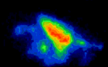
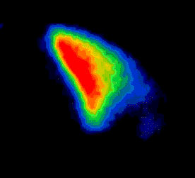
Recently, lung scintigraphy has been adapted to large animal species (Votion et al., 1997) and this sensitive technique has proved to be a unique tool for the detection of subclinical pulmonary dysfunction (Votion et al., 1999a, 1999b). In horses, the regional ventilation of the lung can be visualised with 99mTc-labelled aerosol (Fig. 1). On the other hand, good quality images can not be obtained in calves using aerosol as ventilation imaging agent (Fig. 2). The high respiratory rate found in this species (~ 30 min-1) favours impaction of aerosol in conducting airways and a significant amount of swallowed radioactivity impedes the correct delineation of the lung borders since the forestomachs overlap the caudal part of the lungs. As an alternative to aerosol, inhalation of Technegas has been tried in calves (Fig. 3). Better definition of the ventilated lung was obtained with Technegas because of minimal deposition in conducting airways and decreased activity within the forestomachs (Fig. 4). The decreased contamination of forestomachs may be explained by the difference in radioactivity administration procedure: unsedated calves breathing aerosol for a few minutes tended to salivate and to swallow radioactive contaminated saliva, thus contaminating oesophagus and forestomachs. On the other hand, only a few breaths were necessary to administer the totality of the Technegas dose available in the generator. Therefore, the alimentary tract contamination induced fewer artefacts in the Technegas deposition image.
These preliminary studies demonstrated the feasibility of using Technegas for the visualisation of the ventilated lung in healthy calves breathing spontaneously.
Before using Technegas as a ventilation imaging agent in research studies, the Technegas and 81mKr (i.e. a true gas) distribution patterns must be quantitatively compared to confirm the value of Technegas in ventilation studies especially in abnormal lung function.
BIBLIOGRAPHY
 |
|
Figure 1: 99mTc aerosol ventilation image in horses. Ventilation image of the left lung acquired following inhalation of 99mTc-aerosol (left lung viewed from the side). |
 |
|
Figure 2: 99mTc aerosol ventilation image in calves. In calves, aerosol deposition images show intense central airway impaction and contamination of forestomachs that prevent interpretation of the scans as ventilation images (lateral view of the right lung). |
 |
|
Figure 3: Administration of Technegas for ventilation imaging in calves. The Technegas was delivered to the calves via the Technegas administration tubing system connected to an airtight facemask. |
 |
|
Figure 4: Technegas ventilation images in calves. In healthy calves, Technegas enables high quality images to be obtained since no central deposition of radioactivity is seen. |
|
|
|
|