Experimental
Absorption and fluorescence excitation spectra are observed using an apparatus for studying gas phase photochemistry which was constructed on the beam line BL2A of the Synchrotron Radiation facility (UVSOR) at Institute for Molecular Science (IMS). In brief the focused dispersed radiation from a 1 m Seya-Namioka monochromator enters a photoabsorption gas cell (with a pass length of 12.3 cm) through a LiF window. The transmitted photons are detected by a photomultiplier tube (PMT) after conversion to UV and visible light by means of a light converter, i.e. a sodium salicylate coating on the outer side of the exit LiF window. The fluorescence excitation spectrum of total emission (B−X, C−A and D−X) has been measured by detecting the fluorescence photons emitted and focused in the direction perpendicular to the light beam with a PMT (Hamamatsu R585).
The dispersed fluorescence spectra were recorded using a 32 cm monochromator (Jobin Yvon HR-320) blazed at 250 nm with a resolution of the observed emission. The total photoemission cross section, σem, has been determined by comparing the excimer fluorescence intensity with that for OH(A−X) formed in photodissociative excitation of H2O at 133.57 nm light whose cross section has been determined as σem = 0.29 Mb (10−18 cm2).
In order to determine absolute R values in a wide wavelength range at very many frequency points within a limited measurement time, two methods have been applied, i.e. i) a point-by-point determination of the absolute R value and ii) the measurement of relative fluorescence intensities using two sets of calcite polarizer-photomultiplier detectors which we call a dual-detector system. The former method determines the absolute R values at a few exciting wavelengths and the latter determines the relative intensities for the polarized emissions parallel and perpendicular to the electric vector of the exciting light, a scaling factor of which is estimated by scaling the relative values to the absolute ones determined by the former technique.
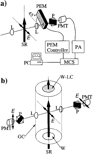
Schematics of an apparatus used for photofragment polarization measurement; a) System for absolute fluorescence anisotropy measurement (PEM-MCS system), b) Experimental set-up to determine the relative ratio of the polarized fluorescence intensity parallel to that perpendicular to electric vector, E, of the incident SR light in a wide wavelength range. GC: gas cell, L: lens, MCS: multichannel scaler, P: polarizer, PA: pulse amplifier, PC: personal computer, PEM: photoelastic modulator, PMT: photomultiplier tube, SR: synchrotron radiation, W: LiF window, W-LC: light converter (sodium salicylate) coating on the outer side of the LiF window.